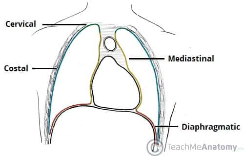
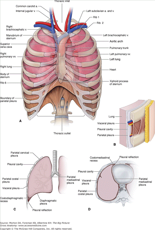
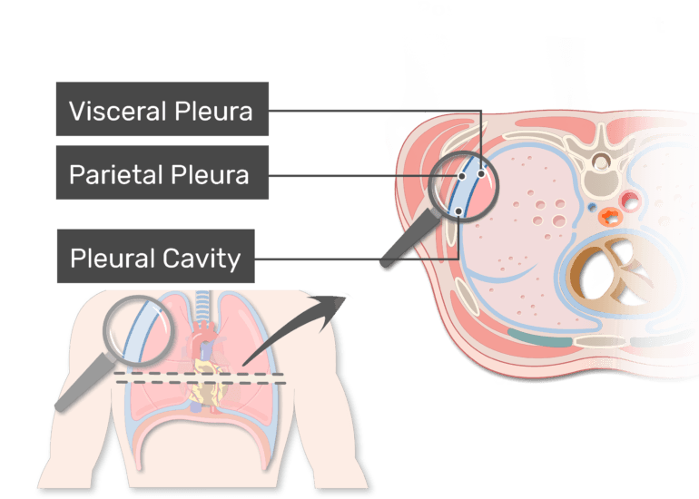
The Space Between the Two Pleural Walls Is Called the
The organisms responsible for the pneumonia may infect the fluid in a pleural effusion known as an empyema. Pleural effusion is a buildup of fluid in the space between the outside lining of your lung.

The Pleurae Visceral Parietal Teachmeanatomy
In addition to answering each question take a moment to appreciate the relative and absolute sizes of the cardiac structures the global and regional function of the right and left ventricles and the appearance of normal valves.

. Another complication is the accumulation of fluid in the space between the lung tissue and the chest wall lining known as a pleural effusion. The walls of your alveoli are normally coated. We would like to show you a description here but the site wont allow us.
This quiz will review basic images and normal anatomy of transthoracic echocardiography.

Deltoid Anatomy Deltoid Is A Large Multipennate Muscle Responsible For The Rounded Contour Of The Sho Anatomy Upper Limb Anatomy Human Anatomy And Physiology

2 Pleura And Lung Flashcards Quizlet

Pleura Anatomy Concise Medical Knowledge

Anterior Compartment Of The Arm Anatomy The Anterior Compartment Of The Arm Contains Three Muscle Arm Anatomy Human Anatomy And Physiology Upper Limb Anatomy

Pleura Space Anatomy Abstract Europe Pmc

Ap 50 10 29 1 Pathology Of Lung 1 Respiratory Therapy Notes Respiratory Therapy Student Nursing School Survival

Pleura Anatomy The Thoracic Cavity Lies Within The Walls Of The Thorax And Is Separated From The Ab Thoracic Cavity Body Anatomy Human Anatomy And Physiology

Chapter 3 Lungs Basicmedical Key

Pleura Space Anatomy Abstract Europe Pmc

Waldeyer S Ring Head Anatomy Boots Anatomy

𝐒𝐭𝐫𝐮𝐜𝐭𝐮𝐫𝐞 𝐚𝐧𝐝 𝐟𝐮𝐧𝐜𝐭𝐢𝐨𝐧 𝐨𝐟 𝐭𝐡𝐞 𝐇𝐞𝐚𝐫𝐭 Nursing Notes Heart Structure Cardiac Cycle

Pleura Or Pleurae And Pleural Cavity Of The Lungs


Comments
Post a Comment